الأربعاء، 27 أبريل 2011

النقرس.. داء لم يعد مقتصرا على الأثرياء


عندما كتب الشاعر والطبيب البريطاني ريتشارد بلاكمور عن النقرس عام 1726، وصفه بأنه «نكبة العظماء والأثرياء وميسوري الحال». وكان يعرف أن هذا الداء يقتصر على الشريحة العليا من المجتمع: إذ
من سواهم يستطيع تناول اللحوم الحمراء المشوية ولحم الطرائد يوميا بعد غمرها في مواد لنقعها؟ وكانت اللوحات الفنية توضح السمات الدالة على مكانة المرء الاجتماعية الراقية من خلال تصويره ممدا على أريكة ورجلاه الملفوفة بالضمادات واللتان تؤلمانه موضوعة على مقعد بلا ظهر.

داء الجميع

* لكن في أميركا، لا ينبغي لك أن تتمتع بمكانة اجتماعية رفيعة حتى تعيش في رخاء، فالجميع بدءا من عمال المصانع إلى الموظفين معرضون للإصابة بداء النقرس. وقد أوضحت أحدث الدراسات زيادة معدل انتشار المرض بمقدار ثلاثة أمثال منذ نهاية السبعينات، حيث يعاني نحو 80 في المائة من الأميركيين من هذا الداء. وتزداد نسبة الإصابة به بين النساء، وهو ما يخالف الاعتقاد السائد.

وترجع زيادة انتشار المرض إلى تغير نمط الحياة، فنحن نعيش في «ظروف سهلة» بمعنى أن أكثرنا يستطيع شراء بروتين حيواني ومشروبات غازية. ويعاني ثلث البالغين الأميركيين تقريبا من السمنة التي تمثل عاملا من عوامل الإصابة بمرض النقرس على حد قول غاري كورهان، اختصاصي الأمراض الوبائية بجامعة هارفارد. وقد أوضح عمله مع هايون تشوي، اختصاصي الأمراض الروماتيزمية، أن تناول مشروبين غازيين أو أكثر من المشروبات المحتوية على عصير الذرة الذي يحتوي على نسبة عالية من الفركتوز يعرض لخطر الإصابة بمرض النقرس.

ومن المفارقة أن يكون ارتفاع متوسط العمر أحد الأسباب الأخرى لمرض النقرس، فبعض العقاقير التي تبقي على حياة الأميركيين رغم إصابتهم بارتفاع ضغط الدم والكلى وما غير ذلك من الأمراض مثل «ناياسين» (فيتامين بي - 3) و«ثيازيد»، ومدرّات البول مثل هيدروكلوروثيازايد والفورسيميد (لازيكس) التي اتضح أنها تزيد مستوى حمض اليوريك في الدم. ويمكن لزيادة حمض اليوريك أن تؤدي إلى الإصابة بمرض النقرس.

ويقول روبرت تيركيلاتوب، رئيس الأمراض الروماتيزمية بقسم الأطباء البيطريين في المركز الطبي بسان دييغو: «كلما طالت مدة تناول العقاقير المدرّة للبول والأنواع الأخرى من الأدوية، زادت نسبة حمض اليوريك في الدم بما يؤدي إلى زيادة احتمالات الإصابة بمرض النقرس».

ومن العوامل الأخرى تزايد اهتمام صناعة الأدوية بالمرض مع تزايد حالات الإصابة به. فمع توفير شركات الأدوية لعقاقير جديدة يتزايد وعي الناس بالمرض وأعراضه على حد قول بيشينس وايت، أستاذ بكلية الطب بجامعة جورج واشنطن ونائب رئيس مؤسسة التهاب المفاصل.

حمض اليوريك

* يعد مرض النقرس مرضين بوجه من الأوجه: الأول هو ارتفاع نسبة حمض اليوريك في الدم الذي يحدث عندما لا يقوم الجسم بتكسير البورينات purines وهي وحدات الحمض النووي التي توجد بكثرة في أنواع معينة من اللحوم والمحار والبيرة. وتحدد سرعة تحلل البورينات وسرعة التخلص من حمض اليوريك خارج الجسم عن طريق الكلى، مستوى حمض اليوريك في الدم. وتختلف هذه العوامل حسب العوامل الوراثية والعقاقير التي نتناولها لعلاج حالات مثل ارتفاع ضغط الدم والسكري وأمراض الكلى.

عندما تتشبع أنسجة بعينها بالحمض، تتشكل أجسام بلورية مثل نوع من السكر لا يذوب تماما في الشاي! ويعتمد التبلور على درجة الحرارة وهو ما يفسر سبب حدوث أكثر من نصف مراحل النقرس الأولى في مفصل إبهام القدم، حيث تقل درجة حرارة أقدامنا عن باقي أجزاء الجسم بمقدار خمس درجات وإصبع إبهام القدم على وجه الخصوص تكون أكثر برودة لأنها أكثر بروزا. كذلك يشير تيركيلاتوب إلى أن هذا المفصل يكون أكثر عرضة لتكون بلورات اليوريات بسبب الضغط الواقع عليه من المشي.

النقرس

* ويستجيب جهاز المناعة لتكون بلورات النقرس في المفاصل ويسمى هذا الالتهاب بالتهاب المفاصل وهو ما يجعل مرض النقرس مؤلما. وفي المراحل المتقدمة من مرض النقرس تتكون البلورات على هيئة كتل بيضاء طبشورية تسمى حصى في كل مكان بالجسم بدءا من أصابع القدم ومفاصل المرفق وصولا إلى أصابع اليد والرسغ والأذن.

ليس كل من يسرف في الشرب والأكل يصاب بمرض النقرس، لكننا نشفق على الذين يفعلون ذلك. ومن بين العظماء الذين أصيبوا بهذا الداء آكلو للحوم الحمراء مثل تشارلز داروين وتوماس جيفرسون وإسحاق نيوتن وهنري الثامن.

ومنذ سنوات قليلة، أخذ علماء التاريخ البشري المستحاثي عينة من خنصر الإمبراطور الروماني تشارلز الخامس الذي كان يعرف عنه إصابته بمرض النقرس وتم الاحتفاظ بها في أحد الأديرة بإسبانيا. واكتشفوا أن إصبعه مليئة ببلورات من اليوريا متآكلة ذات نتوءات متجمعة على شكل حصى في عظامه التي تعود إلى القرن السادس عشر. مسكين تاشرلز، فمملكته كانت ممتدة من الأميركتين إلى المحيط الهندي، لكنه لم يكن يستطع أن يمتطي حصانا! ووصف بلاكمور في كتابه الذي صدر عام 1726 الذين يعانون من مرض النقرس بأنهم «ذوو بشرة زهرية قوية». وبحسب جون فيلدينغ، وهو من أصحاب متجر بقالة «برود برانش ماركت» البالغ من العمر 37 عاما، قد يفسر هذا تهديد المرض لصناعة الأغذية التي تتسبب في الإصابة بمرض النقرس. ويقول فيلدينغ الذي ظهرت عليه أعراض المرض لأول مرة عندما كان في الثلاثين من العمر: «كل طاه يعاني من زيادة في الوزن مصاب بمرض النقرس. لقد تناولت صينية كبيرة من كبد الدجاج وشربت عددا كبيرا من زجاجات البيرة. واستيقظت في منتصف الليل إثر شعور بحرقة في إبهام قدمي اليسرى. وكانت الملاءة تبدو مثل ملاءة من حديد تضغط على قدمي».

لقد كان جدّ فيلدينغ لأمه مصابا بالمرض نفسه الذي يمكن أن يتوارثه أفراد العائلة. وأضاف فيلدينغ الملتحي والذي يخفي 40 رطلا (الرطل يساوي 453 غراما تقريبا) إضافيا كان يتمنى لو لم يكن يحملها: «إنه لهذا السبب فإن المرض مرتبط بالمتعة». وقالت تريسي ستانارد شريكة فيلدينغ في العمل: «هذا هو أنت، شخص منغمس في الملذات».

لكن لا يصح هذا الوصف في كل الأحوال، فبعض المصابين بمرض النقرس تكون أجسامهم متوسطة الحجم. وفي الوقت الذي لا يشيع هذا المرض بين السيدات قبل سن انقطاع الطمث نظرا للحماية التي يوفرها لهم هرمون الأستروجين، ينتشر المرض بين السيدات من كبار السن. ويقول وايت من مؤسسة التهاب المفاصل: «إنك ترى سيدات تجاوزن الخمسين مصابات بمرض النقرس وتزداد النسبة بين السيدات اللاتي تجاوزن الثمانين مقارنة بالرجال لأن السيدات يعشن في الغالب مدة أطول من الرجال».

عقاقير قديمة وجديدة

* أصيبت جيرالدين وايت، موظفة فيدرالية متقاعدة تبلغ من العمر 67 عاما وتعيش في بالتيمور، بمرض النقرس عام 2005 بسبب مرض في الكلى نتج عن علاج لمرض الذئبة. ظلت جيرالدين تعاني لمدة ثلاث سنوات من آلام في أصابع قدميها ويديها ومعصميها وركبتيها.

وفي الواقع فإن عقاقير معالجة النقرس قديمة قدم المرض نفسه. وعُرف أحد تلك العقاقير وهو مسكن مستخرج من الزعفران يسمى «كولشيسين» في القرن الثامن عشر حتى أن بن فرانكلين، الذي كان مصابا بمرض النقرس، كتب عنه أطروحة كاملة. لكن «كولشيسين» colchicine لا يعالج المشكلة الرئيسية. وكانت جيرالدين تعاني حساسية من أكثر العقاقير المعالجة لحمض اليوريك وهو «ألوبيورينول» allopurinol. لكن عام 2006 اشتركت في تجربة على عقار جديد يسمى «بيغلوتيكاس» pegloticase (كريستيكسا Krystexxa) الذي يحفز انحلال حمض اليوريك.

وفي غضون ستة أشهر من بداية العلاج ذابت البلورات المتكونة في يدي ورجلي جيرالدين وأصبحت قادرة على العودة إلى حياتها الطبيعية. لكن يُنصح بتناول عقار «بيغلوتيكاس» إلا في 3 في المائة فقط من حالات الإصابة بالمرض. وثمن العقار باهظ وله أعراض جانبية وليس فعّالا في كثير من الأحيان. لكن ربما يفيد في حالات بعض المرضى الذين لا يستطيعون تناول الألوبيورينول أو العقاقير الشراب الأخرى المستخدمة في علاج حمض اليوريك.

منذ توقف جيرالدين عن تناول عقار «بيغلوتيكاس» ظلت نسبة حمض اليوريك في الدم طبيعية من خلال تناول عقار جديد هو «فيبوكسوستات»febuxostat (ألوريك Uloric). وذُهل هيربرت باراف، طبيب الأمراض الروماتيزمية المعالج لها والمقيم في ويتون، من النتائج التي حققها عقار «بيغلوتيكاس» وزاد عدد المشتركين في تجربة العقار. وقال: «عادة ما يستغرق التخلص من بلورات اليوريات من خمس إلى ست سنوات، لكنه لم يستغرق في حالتها سوى بضعة أشهر».

وهناك عقار آخر في السوق يسمى «كولكريس» Colcrys وهو تركيبة معدلة من «الكولشيسين». لكن يصل ثمن القرص منه إلى 8 دولارات في حين أن ثمن القرص من العقاقير التي تشبهه ولم تعد متوفرة في السوق 6 سنتات فقط. وعادة ما يثير ارتفاع سعر عقار «كولشيسين» قلق بعض مرضى النقرس الذين لا يتمتعون بتأمين صحي بحسب واين غرودي، عالم أمراض وراثية في لوس أنجلوس.

خفض الوزن

* وأدى توفر العقاقير الجديدة وصعوبة اتباع نظام غذائي إلى تغيير الوصفات الطبية التي يعطيها الأطباء لمرضى النقرس. ويقول باراف: «عمليا، لن يكون اتباع نظام غذائي، علاجا ناجعا إلا إذا تم فقدان الوزن بشكل كبير. يتبع البعض نظام غذائي محدد لعلاج النقرس». ويقول باراف أستاذ الطب بجامعة جورج واشنطن لمرضاه الذين تم السيطرة على مستوى حمض اليوريك في دمائهم من خلال تناول العقاقير إنهم يستطيعون تناول ما يريدونه من الأطعمة ما دامت خالية من الكحول. ويقول باراف: «هل تحب السلطعون ومستوى حمض اليوريك في الدم طبيعي؟». وأضاف أن فقدان الوزن طريقة جيدة لعلاج حالات مختلفة، لكن النظام الغذائي القائم على تناول كمية محدودة من البورينات ليس ضروريا لعلاج النقرس.

وتصح كلمات بلاكمور عن الوقاية من مرض النقرس، حيث يقول إن «الاعتدال في الأكل والحد من تناول المشروبات الروحية القوية وممارسة التمرينات الرياضية والحد من الانغماس في الملذات من الوسائل التي تقي من شر هذا المرض».

الاثنين، 25 أبريل 2011

السمنة.. تلحق الضرر بالهرمونات الذكرية والنشاط الجنسي والبروستاتا


لم يعد سرا من الأسرار أن البدانة والسمنة تمثلان مشكلتين كبيرتين في الولايات المتحدة، إذ يحتاج نحو ثلثي الأميركيين اليوم إلى خفض أوزانهم، بينما تزداد أعداد الأطفال والبالغين البدينين بوتيرة
مخيفة.

وليس سرا أن السمنة سيئة لصحة الفرد، فزيادة الدهون في الجسم تزيد من مستويات الكولسترول المنخفض الكثافة (LDL) «الضار»، ومن مستويات الدهون الثلاثية، وتقلل في الوقت نفسه من مستويات الكولسترول العالي الكثافة (HDL) «الحميد». كما تؤدي السمنة إلى الإخلال بقدرة الجسم الطبيعية للاستجابة للأنسولين، الأمر الذي يؤدي إلى زيادة مستويات السكر، والأنسولين، في الدم.

كما أن السمنة تقود إلى أكثر من زيادة هذه الأرقام، إذ إنها تؤدي إلى تدهور الصحة عموما، فهي تزيد من مخاطر النوبة القلبية، والسكتة الدماغية، والإصابة بضغط الدم المرتفع، والسكري، وحصى المرارة، والسرطان، والتهاب المفصل العظمي، وانقطاع التنفس أثناء النوم، وتشحم الكبد، والكآبة.

وإجمالا فإن السمنة حالة قاتلة. وفي الواقع فإن السمنة والخمول مسؤولان عن نحو 1000 وفاة بين الأميركيين يوميا. وإن استمرت الأحوال بهذا الشكل، فإنها ستتخطى التدخين بوصفها من المسببات الرئيسية، التي يمكن تفاديها، للوفاة في الولايات المتحدة.

وتؤثر السمنة على الرجال والنساء بنفس القدر، إلا أن ما يثير الدهشة هو أن الرجال يتحملون جهدا أكبر بسببها، لأن السمنة تلقي بظلالها الكثيفة على الهرمونات الذكرية، وعلى الجنس، وعلى صحة البروستاتا.

هل وزني كبير؟

* نظرة على نفسك في المرآة قد تشير إلى زيادة وزنك، إلا أن المعيار الذهبي المعتمد حاليا هو «مؤشر كتلة الجسم» (body mass index) أو (BMI)، الذي يحسب كحاصل قسمة الوزن بالكيلوغرام على مربع الطول بالمتر. ويمكن استخدام الرابط الإلكتروني التالي لحسابه: www.nhlbisupport.com/bmi .

وتقدم قيم هذا المؤشر تقديرات معقولة لدهون الجسم. فالقيمة التي تقع بين 25 و30 تضع الإنسان في مرتبة البدانة، بينما تشكل القيمة 30 فأكثر مرتبة السمنة. إلا أن هذا المؤشر لا يوفر المعلومات حول كيفية توزيع الدهون. وبينما يعرف العلماء أنه لا توجد فائدة من أي فائض للدهون فإن دهون البطن تعتبر من أشد أنواعها ضررا.

لذا؛ ولكي تتعرف على المخاطر المحدقة بك فعليك أن تقيس ببساطة طول محيط الخصر عبر موقع السرة. وبالنسبة للرجال فإن الخطر يحدث عندما يزيد مقداره على 37.5 بوصة (95.25 سم) بينما تبدأ المشكلات عندما يزيد على 40 بوصة (101.6 سم).

السمنة وهرمون الذكورة

* «التيستوستيرون» (testosterone) هو هرمون الذكورة الرئيسي. وبصفته هذه فإنه مسؤول عن ظهور الصوت العميق، والعضلات الكبيرة، والعظام القوية التي هي من خصائص الذكور، وعن نمو الأعضاء التناسلية الذكرية وإنتاج الحيوانات المنوية (الحيامن)، والشهوة الجنسية، ومسؤول أيضا عن نمو شعر اللحية. وبعد تحوله إلى «ديهيدروتيستوستيرون» (dihydrotestosterone)، فإن هذا الهرمون يحفز على نمو البروستاتا وهي حالة غير مرحب بها للرجال في الأعمار المتقدمة.

وترتفع مستويات «التيستوستيرون» عند مرحلة البلوغ ثم تأخذ في الاستقرار في السنين التالية لها، ثم تبدأ في التناقص عند بدايات أواسط العمر. ولأن الهبوط في مستوى «التيستوستيرون» يكون في المتوسط في حدود 1% سنويا، فإن غالبية الرجال الكبار يحافظون على مستوياته الطبيعية. إلا أن أي أمر يسرع في تدهور هذه المستويات يمكن أن يصيب الرجال بحالة «نقص أو عوز التيستوستيرون» (testosterone deficiency).

السمنة تؤدي إلى تقليل مستويات «التيستوستيرون»، فقد وجدت دراسة نشرت عام 2007 أجريت على 1667 رجلا من عمر 40 فأكثر، أن كل زيادة بدرجة واحدة فقط في مقدار مؤشر كتلة الجسم ارتبطت بتدهور بنسبة 2% في مستويات «التيستوستيرون». وإضافة إلى ذلك فقد وجدت دراسة نشرت عام 2008 أجريت على 1802 من الرجال من عمر 30 سنة فأكثر، أن زيادة محيط الخصر كان مؤشرا أقوى للتنبؤ بتدني مستويات «التيستوستيرون» مقارنة بمؤشر كتلة الجسم. إذ ظهر أن زيادة طول الخصر بـ4 بوصات (10 سم تقريبا) أدت إلى زيادة بنسبة 75% من احتمال تعرض الإنسان إلى انخفاض مستويات «التيستوستيرون»، بينما لا تزد فترة 10 سنوات من الهرم من هذا الاحتمال إلا بنسبة 36%.

وإجمالا فإن طول محيط الخصر كان أقوى مؤشر لمفرده في التنبؤ بظهور حالة نقص «التيستوستيرون».

وإن كنت لا تزال متشككا في نتائج هاتين الدراستين الأميركيتين فعليك التأمل في ما أوردته دراسة أسترالية وجدت أن واحدا من كل سبعة من المعانين من السمنة يمكنهم الاستفادة من العلاج التعويضي بـ«التيستوستيرون»، وهذا المعدل أعلى بأربع مرات من المعدل المماثل لدى الأشخاص الذين لا يعانون من السمنة.

ضعف الانتصاب

* رغم أن الرجال المصابين بضعف الانتصاب erectile dysfunction (ED) يلقون باللوم في مشكلتهم في أغلب الأحيان على «التيستوستيرون»، فإن الاضطرابات الهرمونية لا تتسبب إلا في 3% من حالات ضعف الانتصاب هذا من جهة، ومن جهة أخرى فإنه وحتى ومع وجود مستويات طبيعية من «التيستوستيرون»، فإن الرجال السمان يعانون من تعرضهم لخطر أعلى للإصابة بضعف الانتصاب.

وقد وجدت دراسة لجامعة هارفارد أن الرجل الذي يبلغ طول محيط خصره 42 بوصة (106.7 سم) يتعرض مقدار مرتين أكثر لخطر وقوع هذه المشكلة مقارنة برجل طول محيط خصره 32 بوصة (81.28 سم). كما ربط باحثون برازيليون أيضا بين سمنة البطن وضعف الانتصاب، ولكن فقط للرجال من أعمار 60 سنة فأكثر. وأفادت دراسة من كاليفورنيا بأن قيمة مؤشر كتلة الجسم البالغة 28 (بدانة وليست سمنة) زادت من احتمالات تعرض الرجال بضعف الانتصاب بنسبة تزيد على 90%.

إلا أن تأكيد وجود الصلة بين هذا وذاك أمر، والبحث عن وسائل تحسين وظيفة الانتصاب، أمر آخر. فقد وجدت دراسة من ماساتشوستس أن فقدان الوزن يمكنه بالفعل تحسين الأمور لدى الرجال البدينين المعانين من ضعف الانتصاب. وقد قدم علماء إيطاليون نتائج مشابهة عندما وضعوا عشوائيا 110 من الرجال السمان المصابين بضعف الانتصاب في مجموعة اتبعوا فيها حمية غذائية ومارسوا تمارين رياضية، ومجموعة أخرى تمت العناية بهم صحيا وفق الطرق المتبعة. وبعد سنتين تمكن أكثر من 30% من الرجال في مجموعة الحمية الغذائية والتمارين الرياضية من تصحيح ضعف الانتصاب تماما من دون اللجوء إلى عقاقير طبية، مقابل 6% في مجموعة الرعاية الصحية. وظهر أن الرجال الذين فقدوا أوزانهم أكثر من غيرهم هم الذي حققوا أكثر تلك الفوائد.

الوظيفة التناسلية

* وتلقي السمنة بظلالها الداكنة على الحالة الجنسية، كما قد تؤدي إلى إضعاف الخصوبة. فقد ربط بحث أميركي بين السمنة وتناقص أعداد «الحيامن»، وكذلك قلة حركيتها. وتوصل علماء ألمان إلى نتائج مماثلة لدى رجال بين أعمار 20 و30 سنة.

حصى الكلى

* وإن كان ضعف الانتصاب يؤدي إلى آلام نفسية لدى الرجل وشريكة حياته، فإن حصى الكلى لا تمثل مشكلة شخصية بقدر ما تقود إلى المعاناة من آلام بدنية. وتظهر حصى الكلى لدى الرجال أكثر بمرتين من النساء، وتزيد السمنة من أخطار حدوثها.

وقد وجدت دراسة لجامعة هارفارد لـ45 ألفا و988 من الرجال الذين تراوحت أعمارهم بين 40 و75 سنة، أن قيم مؤشر كتلة الجسد الكبيرة وزيادة طول محيط الخصر ترتبط كل منهما بزيادة خطر ظهور حصى الكلى.

وقد ظهر أن الرجال الذين يزيد وزنهم بأكثر من 39 رطلا (16 كلغم تقريبا) بعد سن الـ20، تزداد لديهم على الأكثر حالات حصى الكلى بنسبة 39% عن غيرهم من الآخرين الرشيقين. أما الرجال الذين يصل وزنهم إلى أكثر من 220 رطلا (100 كلغم تقريبا)، ففقد زادت نسبة احتمال تعرضهم لحدوث حصى الكلى 44% أكثر من الذين يصل وزنهم إلى 150 رطلا (68 كلغم).

وعلى الرغم من أن هذه النتائج الأميركية لم تقدم تفسيرات لأسباب وجود هذه الصلة، فإن دراسات من أوروبا وآسيا أظهرت أن الأشخاص البدينين يعانون من تراكم كميات فائضة من الكالسيوم ومواد كيميائية أخرى في البول، الأمر الذي يؤدي إلى تكوين تلك المواد للحصى.

تضخم البروستاتا الحميد

* يشيع ظهور حالة «تضخم البروستاتا الحميد» Benign prostatic hyperplasia (BPH) مع تقدم عمر الرجال. كما تزداد هذه الحالة مع السمنة.

وقد وجدت دراسة لجامعة هارفارد على 25 ألفا و892 رجلا أن طول محيط الخصر يرتبط بقوة مع خطر ظهور أعراض تضخم البروستاتا الحميد لدى الرجال. فالرجال الذين يبلغ طول خصرهم 43 بوصة (109.2 سم) أو أكثر، كانوا يحتاجون بمعدل 2.4 مرة إلى الخضوع إلى عملية جراحية لعلاج تضخم البروستاتا الحميد، مقارنة بالرجال الذين كان محيط خصرهم يقل عن 35 بوصة (88.9 سم). ولم توظف دراسة هارفارد هذه مؤشر كتلة الجسم كعامل خطر مستقل، وذلك عكس دراسة أخرى أجريت في بالتمور. وقد قدم باحثون من بالتيمور وآخرون من الصين تفسيرات لهذه الصلة، فقد استخدموا تقنيات التصوير بالموجات فوق الصوتية والرنين المغناطيسي لقياس غدة البروستاتا ووجدوا ازديادا في حجم هذه الغدة لدى الأشخاص المصابين بسمنة البطن.

ألغاز البروستاتا

* تشتهر غدة البروستاتا بألغازها وتناقضاتها العلمية. وإليكم واحدا إضافيا منها، إذ تزداد مستويات مولد المضادات الخاص بالبروستاتا prostate-specific antigen (PSA) مع تضخم غدة البروستاتا، وعلى الرغم من أن السمنة تبدو وكأنها تدفع البروستاتا إلى التضخم، فإنها تخفض في الوقت نفسه من مولد المضادات الخاص بالبروستاتا (PSA) هذا.

ووفقا لـ«تقييم الصحة والتغذية الوطني 2001 - 2004» للرجال المتحدرين من أصول قوقازية الذين كانت أعمارهم تبلغ 40 سنة فأكثر، فإن زيادة بمقدار 5 بوصات (12.5 سم تقريبا) في طول محيط الخصر أدت إلى تدهور بنسبة 6.6% في تركيز مولد المضادات الخاص بالبروستاتا (PSA) في الدم.

ولكن، وبخلاف الكثير من ألغاز البروستاتا، فقد أمكن تفسير هذا اللغز، فقد توصلت دراسة على 13 ألفا و634 من المصابين بسرطان البروستاتا إلى أن الرجال الذين يمتلكون مؤشرا أكبر لكتلة الجسم تقل لديهم مستويات مولد المضادات الخاص بالبروستاتا، وهذا لا يحدث لأن البروستاتا تفرز كميات أقل من مولد المضادات هذا، بل لأن السمنة تزيد من حجم الدم، ولذلك فإن تركيز مولد المضادات يقل في الدم.

سرطان البروستاتا

* ولأن السمنة تقلل من تركيز مولد المضادات الخاص بالبروستاتا (PSA)، فإن الأطباء يجدون صعوبة أكبر في توظيف قياسات هذا المؤشر، أي (PSA)، في رصد سرطان البروستاتا لدى الرجال البدينين.

وتؤثر السمنة على الجوانب البيولوجية الممهدة لسرطان البروستاتا، فقد أشارت أبحاث أجريت في مناطق مختلفة من العالم إلى أن الدهون الزائدة في الجسم تزيد من خطر تعرض الرجال لسرطان البروستاتا. وأظهرت دراسة لجمعية السرطان الأميركية على 404 آلاف و576 رجلا هذه الصلة، إذ تبين أن البدانة تزيد من الخطر على الرجال بنسبة 8%، بينما تزيد السمنة من ذلك الخطر بنسبة 20%، وأخيرا فإن الخطر يزداد بسبب السمنة الفائقة إلى 34%.

ولم يتوقف الأمر عند هذا الحد، فقد ظهر أن السمنة تزيد من احتمال انتشار سرطان البروستاتا إلى خارج هذه الغدة، وأنها تزيد من احتمال عودة السرطان بعد معالجته. وإضافة إلى هذا فإن السمنة تزيد أيضا من حالات سلس البول بعد إجراء عمليات جراحية لإزالة سرطان البروستاتا.

لماذا تعتبر السمنة مهددة بهذه الدرجة لخطر سرطان البروستاتا؟ لأن الأشخاص البدينين لا يأبهون لطلب المشورة الطبية لامتلاكهم مستويات أقل من مولد المضادات الخاص بالبروستاتا (PSA)، ولذلك فإن تأخر التشخيص هو أحد تلك التفسيرات لأخطارها. كما أن السمنة تغير أيضا من عملية التمثيل الغذائي (الأيض) للهرمونات الجنسية، الأمر الذي يؤثر على نمو أورام سرطان البروستاتا. ولكن الأهم من كل ذلك، ربما، هو أن السمنة تزيد من إنتاج الجسم لعوامل النمو مثل الأنسولين، وعامل النمو الشبيه بالأنسولين insulin-like growth factor 1 (IGF-1). ويزيد كلا هذين العاملين من معدل تكاثر الخلايا، كما أن ازدياد مستويات IGF-1)) في الدم يرتبط بزيادة خطر سرطان البروستاتا، وسرطان القولون، والأورام الخبيثة الأخرى.

قد تبدو السمنة من علامات العصر إلا أنها ظاهرة غير طبيعية، تؤدي إلى حدوث مختلف الأمراض. ولذا؛ فعليك البدء في خفض وزنك اليوم قبل الغد.

دراسة طبية تربط بين مضادات الاكتئاب ومخاطر على القلب


نيو اورليانز (رويترز) - كشفت دراسة أن الرجال متوسطي العمر الذين يستخدمون مضادات للاكتئاب أكثر عرضة للاصابة بضيق في الاوعية الدموية وهو ما يزيد من احتمالات الاصابة بأزمات قلبية وسكتات
دماغية مقارنة بغيرهم الذين لا يتناولون هذه العقاقير.

واظهرت دراسة على عدد من التوائم أدلة على تصلب للشرايين في قياس السمك الداخلي للشريان السباتي بصرف النظر عن نوع مضاد الاكتئاب المستخدم.

واكتشفت الدراسة ان استخدام مضادات الاكتئاب تسبب زيادة قدرها 37 ميكرون في سمك الشريان السباتي او نحو خمسة في المئة. وشملت الدراسة 500 توأم من الرجال متوسط اعمارهم 55 عاما وعرضت في الاجتماع العلمي لطب القلب الامريكي في نيو اورليانز.

وبين كل توأم من جملة 59 توأما يأخذ احدهما مضاد للاكتئاب دون الاخر. واظهر البحث ان الذي يتناول العقار يزيد لديه سمك البطانة الداخلية للشريان السباتي بمتوسط 41 ميكرون.

وبم انه في كل عام يزيد سمك الشريان بعشرة ميكرونات تكون شرايين الشقيق الذي تناول العقار اكبر بأربعة أعوام عن نظيرتها لدى توأمه.

وكانت دراسات سابقة ربطت بين الاكتئاب وزيادة مخاطر الاصابة بأمراض القلب ولكن الدراسة لم تعتبر الاكتئاب دليلا قويا على تصلب الشرايين.

وقال الدكتور اميت شاه زميل طب القلب في جامعة ايموري في اتلانتا الذي عرض الدراسة "لاننا لم نر علاقة بين الاكتئاب في حد ذاته وضيق الشريان السباتي فذلك يدعم حجة انه من المرجح جدا ان مضادات الاكتئاب وليس الاكتئاب الفعلي قد يكون سبب هذه العلاقة."

وأضاف شاه "تذكرنا الدراسة بان الادوية يكون لها في الغالب اثار جانبية لا نشعر بها ويتعين علينا دوما ان نأخذ ذلك في الاعتبار. تقدم هذه العقاقير الكثير من الفوائد ولكن يتعين التعامل مع كل حالة على حدة."

ورجح شاه ان تكون زيادة مستويات مواد كيميائية بعينها في المخ مثل السيروتونين والبافراز نتيجة استخدام مضادات الاكتئاب قد تسبب انقباض الاوعية الدموية مما يؤدي الى نقص في تدفق الدم الى الاعضاء وارتفاع الضغط وهو ما يزيد من خطورة الاصابة بتصلب الشرايين.

وقال شاه "لانها كانت دراسة على توائم فقد اجرينا تحليلا دقيقا يقارن بين أشقاء متشابيهن جينيا بأي حال من الاحوال بنسبة تترواح ما بين 50 الى مئة في المئة ونشأوا في نفس البيئة."

صحة المخ.. تبدأ من الأمعاء


«أحاديث متبادلة» تتم داخل أجسامنا بين البكتيريا المعوية والخلايا المخية، وتلعب دورا مهما في حدوث الاعتلالات النفسية والوظائف المخية. وأكدت النتائج تأثر الجينات المتعلقة بالتعلم والذاكرة في خلايا منطقة «هيبوكامبس
Hippocampus» بالمخ في نموذج فأر تجارب خال من الجراثيم المعوية Germ Free Mice مقارنة بالفئران الطبيعية. كما أكدت العلاقة بين اختلال التوازن البكتيري بالأمعاء وحدوث التوتر وأحيانا الاكتئاب نتيجة اختلال عمل الخلايا العصبية بالمخ.

تحتوي أمعاء البشر على عدة تريليونات من البكتيريا غير الضارة يطلق عليها «البكتيريا الصديقة» أو البكتيريا التكافلية، وهناك شبه إجماع بين العلماء على أن صحة الإنسان وحيويته ترتبط بدرجة كبيرة بما تحتويه الأمعاء من ميكروبات صديقة. لذلك ينصح خبراء الصحة بضرورة المحافظة على هذه الثروة من البكتيريا التي تعيش وتتكاثر في توازن وتكامل يلعب دورا مهما في حماية الجسم من الآفات التي يمكن أن تصيبه من جراء اختلال هذا التوازن.

إن صحة الأمعاء لا تعني فقط التخلص من الغازات والانتفاخ ومضايقات عسر الهضم، فالأبحاث الحديثة تؤكد أن اعتلال صحة الأمعاء يمكن أن يعيث فسادا في الجسم كله، فيؤدي إلى اضطرابات المزاج والتوحد والخرف والإجهاد المزمن والتشحم الكبدي والسمنة والتهاب المفاصل واعتلال المناعة والطفح الجلدي والسرطان وغيرها.

* «حوارات» المخ والأمعاء

* أشارت دراسة نشرت خلال شهر مارس (آذار) 2011 في دورية Neurogastroenterology and motility أجراها باحثو الطب النفسي والعلوم السلوكية بجامعة ماكماستر الكندية، إلى وجود «أحاديث متبادلة» تتم داخل أجسامنا بين البكتيريا المعوية وخلايا المخ، وتلعب دورا مهما في حدوث الاعتلالات النفسية والوظائف المخية.

وأكدت النتائج تأثر الجينات المتعلقة بالتعلم والذاكرة في خلايا منطقة «هيبوكامبس Hippocampus» بالمخ في نموذج فأر تجارب خال من الجراثيم المعوية Germ Free Mice مقارنة بالفئران الطبيعية. كما أكدت العلاقة بين اختلال التوازن البكتيري بالأمعاء وحدوث التوتر وأحيانا الاكتئاب نتيجة اختلال عمل الخلايا العصبية بالمخ.

ويأمل الباحثون في إيجاد مستهدفات جديدة مثل بكتيريا الأمعاء لإيجاد علاجات حديثة خالية من الآثار الجانبية، تستهدف الجسد وتؤثر بشكل فعال على وظائف المخ، وتكون بديلة عن العلاجات الحالية لعلاج الاعتلالات النفسية والتي يصاحبها العديد من الآثار الجانبية.

كما أكد الباحثون أن الموجة المستقبلية من الأبحاث العلمية تتطلع إلى دراسة المزيد عن العلاقة المزدوجة بين المخ والجراثيم المعوية وتأثير هذه العلاقة على الاضطرابات الأيضية، مثل الإصابة بالسمنة ومرض السكري.

* البكتيريا المعوية والتوحد

* وأكدت نتائج دراسة نشرت خلال شهر يناير (كانون الثاني) 2011 في دورية المعهد القومي الأميركي للعلوم، أجراها باحثون بجامعة غرب أونتاريو الكندية، أن اضطرابات الجهاز الهضمي المصاحبة لمرض التوحد Autism ليست مجرد أعراض مصاحبة للمرض، لكنها قد تكون سببا وراء حدوث المرض ذاته.

أجريت الدراسة على مجموعتين من الفئران، المجموعة الأولى خالية من الجرثومات المعوية والثانية تحتوي على الجرثومات المعوية الطبيعية. ولاحظ الباحثون أن المجموعة الأولى كانت أكثر نشاطا وإقبالا على الأنشطة المحفوفة بالمخاطر، وأكدت فحوصات المقارنة بين المجموعتين أن المجموعة الأولى الخالية من الجراثيم المعوية عانت من حدوث تغيرات جينية متعلقة بإفراز الناقلات العصبية (نور أدرينالين - دوبامين - سيروتونين)، مما أدى إلى حدوث اضطرابات السلوك والإصابة بأعراض مرض التوحد.

كما أرجع الباحثون ارتباط البكتيريا المعوية بالاضطرابات السلوكية المصاحبة لمرض التوحد إلى ارتفاع مستويات حمض بروبيونيك Propionic acid، الذي تفرزه البكتيريا المعوية الضارة «كوليستريديا Clostridia» في مجرى الدم ليصل إلى الخلايا العصبية بالمخ ويتسبب في حدوث العديد من الأعراض المميزة لمرض التوحد، مثل فرط النشاط الحركي والميل إلى تكرار الحركات لا إراديا وعدم الاكتراث الاجتماعي.

والجدير بالذكر أن الدراسات السابقة أكدت أن نحو 70 في المائة من أطفال التوحد يعانون من اضطرابات بالجهاز الهضمي، تتمثل في نوبات متعاقبة من الإسهال والإمساك والانتفاخ وألم البطن. كما أشارت بعض الدراسات إلى احتواء أمعاء أطفال التوحد على تركيزات عالية من بكتيريا «كوليستريديا» الضارة (المسؤولة عن إفراز سموم عصبية)، وأكدت دراسات أخرى اختلاف أعداد وأنواع البكتيريا المعوية لدى أطفال التوحد مقارنة بأقرانهم الطبيعيين.

* البكتيريا والتصلب المتعدد

* وأشارت دراسة نشرت خلال شهر يوليو (تموز) 2010 في دورية PNAS لعلماء البيولوجيا في معهد كاليفورنيا للتكنولوجيا Caltech إلى العلاقة بين التغييرات التركيبية للبكتيريا التكافلية بالأمعاء والإصابة بمرض التصلب اللويحي المتعدد MS (أحد أمراض المناعة الذاتية، غير معروفة السبب، الذي يصيب مناطق متعددة بالمخ والنخاع الشوكي).

واكتشف الباحثون أن نموذج فأر التجارب الخالي من الجراثيم لا يصاب بالتصلب اللويحي المتعدد. وأدى إدخال أحد أنواع البكتيريا المعوية Segmented filamentous bacteria، إلى حدوث سلسلة من التفاعلات الالتهابية بالأمعاء، نتيجة تنشيط إفراز خلايا مناعية تسمىTh17، مما أدى إلى حدوث تغيرات مناعية تتسبب في حدوث مرض التصلب اللويحي المتعدد.

والجدير بالذكر أن العديد من الدراسات السابقة أشارت إلى العلاقة بين العدوى الفيروسية أو البكتيرية وحدوث مرض التصلب اللويحي المتعدد أو زيادة أعراضه، لذلك يأمل الباحثون في إجراء المزيد من الأبحاث لدراسة إمكانية استخدام البروبايوتك في الوقاية والتخفيف من حدة أعراض المرض.

* التوتر واختلال البكتيريا

* كما أكدت دراسة نشرت خلال شهر مارس 2011 في دورية Brain، Behavior and immunity أجراها باحثون بجامعة أوهايو الأميركية، أن التعرض للتوتر المزمن يؤدي إلى حدوث تغيرات في التوازن البكتيري بالأمعاء فتصبح أقل عددا وانتشارا، وتغلب عليها البكتيريا الضارة مثل بكتيريا «كوليستريديا». وأكد الباحثون أن هذه التغيرات قد تكون بمثابة مفتاح سر العلاقة بين التوتر المزمن واضطرابات الاستجابة المناعية بالجسم.

القهوة بعد الوجبات السريعة.. ترفع مستوى السكر في الدم


لا يعد تناول وجبة سريعة ممتلئة بالدهون أمرا جيدا، ولكن احتساء كوب من القهوة عقب هذه الوجبة أكثر سوءا، وفقا لما أفادت به دراسة جديدة لجامعة غيلف الكندية University of
Guelph.

* دور الدهون والكافيين

* اكتشفت الباحثة ماري سوليل بيدوان أن مستويات السكر في دم الشخص السليم ترتفع بعد تناول وجبة بها مقدار كبير من الدهون، إلا أن ارتفاع هذه المستويات يتضاعف بعد تناول الوجبة كثيرة الدهون وبعدها قهوة تحتوي على الكافيين، لتصل إلى مستويات شبيهة بتلك الخاصة بالأفراد المعرضين للإصابة بمرض السكري. وتقول بيدوان، التي تسعى للحصول على درجة الدكتوراه وأجرت الدراسة مع الأستاذة بجامعة غيلف ليندساي روبنسون وتيري غراهام: «تخبرنا النتائج أن الدهون المشبعة تتداخل في قدرة الجسم على إزالة السكر من الدم، وعندما يصحب ذلك تناول قهوة محتوية على الكافيين، يكون الأثر أشد سوءا». وأضافت أن «بقاء السكر في دمائنا لفترات طويلة غير صحي لأنه يمكن أن يؤثر على أعضاء جسدنا».

نشرت الدراسة في الأول من أبريل (نيسان) الحالي في مجلة «جورنال أوف نوتريشن» المعنية بالتغذية، وتعد الأولى التي تفحص آثار الدهون المشبعة والقهوة ذات الكافيين على معدلات سكر الدم من خلال توظيف الباحثين لـ«كوكتيل» دهون جديد يحتوي على اللبيدات فقط. وقد شكل ذلك شرابا خاصا سمح للباحثين أن يحاكوا بدقة ما يحدث للجسم عندما يتناول الإنسان الدهون.

* «كوكتيل» تجريبي

* خلال الدراسة شرب رجال أصحاء نحو غرام واحد من الشراب مقابل كل كيلوغرام من وزن الجسم للوجبة الأولى. وبعد ست ساعات حصلوا على وجبة ثانية صغيرة تتكون من شراب سكر.

وفي المعتاد عندما نتناول السكر، يفرز الجسم الأنسولين، الذي يحول السكر بعيدا عن الدم ويوزعه على عضلاتنا، بحسب ما تذكر بيدوان. ولكن الباحثين اكتشفوا أن الوجبة كثيرة الدهون تؤثر في قدرة الجسم على إزالة السكر من الدم. وكانت معدلات السكر في دم الأشخاص محل الدراسة أعلى بنسبة 32 في المائة بالمقارنة بالمعدلات عندما لم يتناول الرجال شرابا، أي «كوكتيل» الدهون هذا. وقام الباحثون أيضا باختبار أثر القهوة المحتوية على كافيين مع الوجبة المحتوية على دهون. ومن أجل هذا الاختبار، حصل المشاركون على ما يساوي كوبين من القهوة المحتوية على كافيين بعد خمس ساعات من تناول الشراب الدهني. ثم أعطوا بعد ساعة شراب سكري. وأظهرت النتائج أن معدلات السكر في الدم ارتفعت بنسبة 65 في المائة بالمقارنة مع معدلاتها عندما لم يتناول المشاركون الدهون أو القهوة المحتوية على كافيين. وتقول بيدوان: «يظهر ذلك أن آثار الوجبة المحتوية على قدر كبير من الدهون يمكن أن تستمر لأربع ساعات. وما تتناوله في الغداء يمكن أن يؤثر على كيفية استجابة الجسم للطعام في وقت لاحق في النهار».

* التأثير على الجهاز الهضمي

* وإلى جانب اختبار معدلات السكر في دم المشاركين، نظر الباحثون في آثار الجهاز الهضمي من خلال قياس هرمون (incretin) الذي يخرج من القناة الهضمية بعد تناول الدهون. وتبعث هذه الهرمونات بإشارات إلى البنكرياس كي ينتج الأنسولين للمساعدة على تخليص الدم من السكر. واكتشف الباحثون أن استجابات هذه الهرمونات للكربوهيدرات تقل بعد تناول الشراب المحتوي على دهون. وتقول بيدوان: «في النهاية اكتشفنا أن الدهون والقهوة المحتوية على كافيين يلحقان الضرر بعملية التواصل بين القناة الهضمية والبنكرياس، مما قد يلعب دورا في عدم قدرة المشاركين على إزالة السكر من دمائهم بسهولة»، كما نقل عنها موقع «ميديكال نيوز توداي» الإخباري الطبي.

وتحظى نتائج هذه الدراسة بأهمية خاصة للأشخاص المعرضين للإصابة بأمراض الأيض (التمثيل الغذائي) ومرض السكري من النوع الثاني، بحسب ما ذكرت. وأضافت: «عرفنا منذ أعوام أن الأشخاص الذين يعانون من النوع الثاني من مرض السكري أو يحتمل أصابتهم به يجب أن يحدوا من تناولهم للكافيين. ويعد احتساء القهوة منزوعة الكافيين بدلا من المحتوية على كافيين وسيلة من أجل تحسين قبول الجسم للغلوكوز. ومن المفيد أيضا الحد من تناول الأحماض المشبعة بالدهون الموجودة في اللحوم الحمراء والأطعمة المعالجة والوجبات السريعة. وقد أظهرت هذه الدراسة أن تأثيرات هذه الأطعمة قد يكون خطيرا ويستمر لوقت طويل».

المشي10 آلاف خطوة يوميا.. هل هو ضروري لكبار السن؟


شترت لي ابنتي جهازا لتعداد الخطوات، وطلبت مني أن أمشي 10 آلاف خطوة يوميا. وعندما ارتديت هذا الجهاز لفترة ما، عرفت أني لا أمشي أكثر من 3 آلاف خطوة في
اليوم. هل المشي 10 آلاف خطوة في اليوم هدف واقعي لشخص مثلي يبلغ عمره 70 سنة؟

إن كنت من الأشخاص الأصحاء عموما، فإن المشي 10 آلاف خطوة هو هدف جيد لك. ويعادل هذا العدد من الخطوات المشي ما بين ميلين وثلاثة أميال في اليوم (ثلاثة إلى 5 كيلومترات).

ويمكنك قطع هذه المسافة في حدود 45 دقيقة أو ما يقاربها، وبهذا تحصل على الـ10 آلاف خطوة هذه في اليوم حتى وإن تمتعت بالراحة طيلة بقية اليوم. إلا أن المشي 10 آلاف خطوة لا يعتبر هدفا للأشخاص المصابين بالتهاب المفاصل، وعجز القلب، أو أية مشكلات صحية أخرى.

ولا يمكن لأي شخص القول إنه يجب عليك أن تحقق هذا الهدف كل يوم، ولكن إن كنت قد مشيت عددا قليلا من الخطوات يوما أو يومين، فعليك المشي أكثر فأكثر في الأيام اللاحقة.

cancer breast


الخميس، 21 أبريل 2011

Circle of Willis


Overview
The circle of Willis (circulus arteriosus cerebri) is an anastomotic system of arteries that sits at the base of the brain. The “circle” was named after Thomas Willis by his student Richard Lower. Willis was the author of Cerebri Anatome, a book that described and depicted this vascular ring. Although such a vascular ring had been described earlier, the name Willis has been eponymously propagated.

The circle of Willis encircles the stalk of the pituitary gland and provides important communications between the blood supply of the forebrain and hindbrain (ie, between the internal carotid and vertebrobasilar systems following obliteration of primitive embryonic connections). A complete circle of Willis is present in most individuals, although a well-developed communication between each of its parts is identified in less than one half of the population.
The circle of Willis is formed when the internal carotid artery enters the cranial cavity, bilaterally, and divides into the anterior cerebral artery (ACA) and middle cerebral artery (MCA). The anterior cerebral arteries are then united by an anterior communicating artery (ACOM). These connections form the anterior half (anterior circulation) of the circle of Willis. Posteriorly, the basilar artery, formed by the left and right vertebral arteries, branches into a left and right posterior cerebral artery (PCA), forming the posterior circulation. The posterior cerebral arteries complete the circle of Willis by joining the internal carotid system anteriorly via the posterior communicating arteries (PCOM).
The images below depict the circle of Willis.

Gross Anatomy
Anterior Cerebral Artery
A1 segment and anterior communicating artery
The A1 segment extends from the internal carotid artery (ICA) bifurcation in a medial and superior direction to its junction with the anterior communicating artery (ACOM) within the longitudinal fissure. Branches include the medial lenticulostriate arteries (A1) that supply the anterior hypothalamus, anterior commissure, fornix, striatum, optic chiasm, and optic nerves. ACOM branches include perforators that supply the hypothalamus and optic chiasm.

A2 segment
This portion of the anterior cerebral artery extends from the ACOM artery to its division into the pericallosal and callosomarginal arteries, at the genu of the corpus callosum. Branches include perforators to the frontal lobe, and the recurrent artery of Heubner, which is a large lenticulostriate vessel. This latter vessel supplies the caudate nucleus, internal capsule, and putamen. Other branches of A2 include the orbitofrontal and frontopolar arteries.

A3 segment
These include all branches of the anterior cerebral artery (ACA) distal to the origin of the pericallosal and callosomarginal arteries, but other subdivisions have been used. Many anastomoses occur with distal branches of the middle cerebral artery (MCA) and posterior cerebral artery (PCA).[2] The pericallosal artery travels posteriorly over the corpus callosum and anastomoses with the splenial artery. The callosomarginal artery courses over the cingulate gyrus. A paracentral artery arises from the pericallosal or callosomarginal arteries and supplies the paracentral lobule. This segment terminates by providing parietal arteries to the corpus callosum and precuneus.

Middle Cerebral Artery
Most classification schemes divide the middle cerebral artery (MCA) into 4 segments, including M1 from the ICA to the bifurcation (or trifurcation), M2 from the MCA bifurcation to the circular sulcus of the insula, M3 from the circular sulcus to the superficial aspect of the Sylvian fissure, and M4, which are cortical branches.

M1 segment
Most anatomic studies define the M1 segment as ending where the MCA branches take a right angled turn within the Sylvian fissure; however, the division point of the MCA trunk is considered by most clinicians to be the M1/M2 junction.[3] The MCA most commonly bifurcates but may also trifurcate or quadfurcate.[4] Branches include lenticulostriate arteries that supply the anterior commissure, internal capsule, caudate nucleus, putamen, globus pallidus, and an anterior temporal artery that supplies the anterior temporal lobe.[5]

M2 segment
The M2 segment extends from the main division point of the M1 segment, over the insula within the Sylvian fissure, and terminates at the margin of the insula.

M3 segment
The M3 segment begins at the circular sulcus of the insula and ends at the surface of the Sylvian fissure. This part travels over the surface of the frontal and temporal operculae to reach the external surface of the Sylvian fissure. The M3 and M2 segments give rise to stem arteries from which cortical branches are derived.

M4 segment
The M4 segment begins at the surface of the Sylvian fissure and extends over the surface of the cerebral hemisphere. Its cortical branches supply the frontal, parietal, temporal, and occipital lobes. These branches include orbitofrontal, prefrontal, precentral, central, anterior, and posterior parietal, angular, temporo-occipital, temporal, and temporopolar branches. The MCA branches that form the so-called "candelabra" are the prefrontal, precentral, and central arteries.
 
Posterior Cerebral Artery
A commonly used subdivision for this vessel includes dividing it into a P1 segment from the basilar artery bifurcation to the junction with the posterior communicating arteries (PCOM) artery, a P2 segment from the PCOM artery to the posterior aspect of the midbrain, a P3 segment from the posterior aspect of the midbrain to the calcarine fissure, and a P4 segment that describes terminal branches of the PCA distal to the anterior aspect of the calcarine fissure.

P1 segment andposterior communicating arteries
The P1 segment supplies perforating branches to the brainstem. These are termed the posterior thalamoperforators to distinguish them from the anterior thalamoperforators, which arise from the PCOM artery. The direct perforators supply the thalamus, brainstem, and internal capsule. Short and long circumflex arteries supply the thalamus and midbrain. A meningeal branch may supply the inferior surface of the tentorium cerebelli.[7]

P2 segment
The P2 segment begins at the PCOM artery junction and travels around the lateral aspect of the midbrain. Direct perforators supply the thalamus, internal capsule, and the optic tract. Branches include the posteromedial choroidal artery that supplies the midbrain, pineal gland, thalamus, and medial geniculate body and the posterolateral choroidal artery that supplies the choroid plexus, thalamus, geniculate bodies, fornix, cerebral peduncle, pineal body, corpus callosum, tegmentum, and temporal occipital cortex. A hippocampal artery may be present. The inferior temporal arteries anastomose with anterior temporal branches of the MCA. The parieto-occipital artery arises as a single trunk from the P2 segment more commonly than from the P3 segment. This artery supplies the posterior parasagittal region, cuneus, precuneus, and lateral occipital gyrus.

P3 segment
The P3 segment extends from the tectum to the anterior aspect of the calcarine fissure. The PCA often divides into its 2 terminal branches, the calcarine and parieto-occipital arteries.

P4 segment
The P4 segment begins at the anterior limit of the calcarine fissure and often includes one of the 2 main terminal branches of the PCA, the calcarine artery. The other main terminal branch of the PCA, the parieto-occipital artery, frequently arises from the P2 or P3 segment. The splenial artery arises from the parieto-occipital artery in most individuals and usually anastomoses with the pericallosal artery.
 
Basilar Artery
The basilar artery originates at the junction between the left and right vertebral arteries and travels anterior to the brainstem. Branches include the superior cerebellar artery and anterior inferior cerebellar artery (AICA).[9] The superior cerebellar artery (SCA) arises from the basilar artery immediately prior to the basilar bifurcation. The SCA often comes into contact with the trigeminal nerve and is usually the target of surgical microvascular decompression for trigeminal neuralgia.

The artery sends branches to the tectum, vermis, and the medial aspect of the cerebellar hemisphere. AICA travels toward the cerebellopontine angle. The posterior inferior cerebellar artery (PICA) is the largest of the cerebellar arteries and arises from the vertebral artery. It supplies the medulla, cerebellar tonsils and vermis, and inferolateral cerebellar hemisphere.

Natural Variants
Anterior circulation
The anterior cerebral arteries may be united in a single trunk, which runs in the longitudinal fissure, giving branches to both hemispheres. The left and right A1 segments are asymmetric in size in most individuals and may be absent or fenestrated. Rarely, this segment may travel inferior to or through the optic nerve. An accessory ACA may be present, and the A1 segment may arise from the cavernous or contralateral ICA.

Either the right and left anterior cerebral arteries may run as one vessel (azygos) to divide distally, or one may be a branch of the contralateral artery. Other variations of the ACOM include aplasia, fenestration, and duplication.[10] This vessel may be curved, kinked, or tortuous. The artery is rarely absent. One A2 segment may be hypoplastic; thus, the contralateral A2 supplies both hemispheres. A2 may be duplicated. In an azygos ACA, both A1 segments join to form a single A2 segment. Branches to the contralateral hemisphere may be found.

Posterior circulation
When a fetal PCOM artery is present, the ipsilateral P1 is typically hypoplastic. Variations of the P1 segment include duplication, fenestration, and a bilateral shared origin of the PCA and SCA. A prominent perforating branch may supply portions of both the ipsilateral and contralateral thalamus and potentially the mid brain. The posterior cerebral may course below, rather than over, the oculomotor nerve, or it may be absent and replaced by an accessory contralateral vessel. The posterior cerebral may give rise to the anterior cerebral artery. The posterior cerebral artery may arise from the internal carotid.

The PCOM may be absent, or the branch representing it may fail to join the posterior cerebral. Fenestration of the basilar artery is found in less than 1% of cases. The basilar artery may exist as 2 longitudinal trunks united across the midline. The SCA may be duplicated or absent. The internal auditory artery is most often a branch of the AICA but may arise from the SCA (in up to 25% of cases) or the basilar artery (in less than 20% of cases).

Pathophysiological Variants
Asymmetry of the circle of Willis results in significant asymmetry of flow and is an important factor in the development of intracranial aneurysms and ischemic stroke.[6] Patients with aneurysms are more likely to have asymmetry or an anomaly of the circle, and the presence of a nonfunctional anterior collateral pathway in the circle of Willis in patients with ICA occlusive disease is strongly associated with ischemic stroke. Uncommonly, persistence of fetal anastomoses is found involving the circle of Willis. These include persistent trigeminal, otic, hypoglossal and proatlantal arteries. These arteries, more or less, unite the internal carotid and vertebrobasilar systems. 


الثلاثاء، 19 أبريل 2011

Gallbladder Surgery for Gallstones


B-Lynch surgery technique septate uterus


Hysterectomy


Liver Transplants and Hepatitis C


Quick Linker - 3 - (Liver) Donor Operation


الاثنين، 18 أبريل 2011

Breast Anatomy

Anatomy of the Breast
As with all surgical procedures, understanding the anatomy is crucial prior to performing an operative procedure. Comprehension of breast anatomy enhances the surgeon's ability to perform surgery safely and effectively. For information on various breast surgery procedures, please see the Breast section of eMedicine's Plastic Surgery journal.
Vascular Anatomy
The blood supply to the breast skin depends on the subdermal plexus, which is in communication with underlying deeper vessels supplying the breast parenchyma. The blood supply is derived from (1) the internal mammary perforators (most notably the second to fifth perforators), (2) the thoracoacromial artery, (3) the vessels to serratus anterior, (4) the lateral thoracic artery, and (5) the terminal branches of the third to eighth intercostal perforators. The superomedial perforator supply from the internal mammary vessels is particularly robust and accounts for some 60% of the total breast blood supply. This rich blood supply allows for various reduction techniques, ensuring the viability of the skin flaps after surgery.

Innervation of the Breast
Sensory innervation of the breast is dermatomal in nature. It is mainly derived from the anterolateral and anteromedial branches of thoracic intercostal nerves T3-T5. Supraclavicular nerves from the lower fibers of the cervical plexus also provide innervation to the upper and lateral portions of the breast. Researchers believe sensation to the nipple derives largely from the lateral cutaneous branch of T4.

Breast Parenchyma and Support Structures
The breast is made up of both fatty tissue and glandular milk-producing tissues. The ratio of fatty tissue to glandular tissue varies among individuals. In addition, with the onset of menopause (ie, decrease in estrogen levels), the relative amount of fatty tissue increases as the glandular tissue diminishes.

The base of the breast overlies the pectoralis major muscle between the second and sixth ribs in the nonptotic state. The gland is anchored to the pectoralis major fascia by the suspensory ligaments first described by Astley Cooper in 1840. These ligaments run throughout the breast tissue parenchyma from the deep fascia beneath the breast and attach to the dermis of the skin. Since they are not taut, they allow for the natural motion of the breast. These ligaments relax with age and time, eventually resulting in breast ptosis. The lower pole of the breast is fuller than the upper pole. The tail of Spence extends obliquely up into the medial wall of the axilla.[1]

The breast overlies the pectoralis major muscle as well as the uppermost portion of the rectus abdominis muscle inferomedially. The nipple should lie above the inframammary crease and is usually level with the fourth rib and just lateral to the midclavicular line. The average nipple – to – sternal notch measurement in a youthful, well-developed breast is 21-22 cm; an equilateral triangle formed between the nipples and sternal notch measures an average of 21 cm per side.


Musculature Related to the Breast
The breast lies over the musculature that encases the chest wall. The muscles involved include the pectoralis major, serratus anterior, external oblique, and rectus abdominus fascia. The blood supply that provides circulation to these muscles then perforates through to the breast parenchyma, thus also supplying blood to the breast. By maintaining continuity with the underlying musculature, the breast tissue remains richly perfused, thus preventing complications arising from aesthetic or reconstructive surgery that requires the placement of a breast implant. (For more information, see the Breast section of eMedicine's Plastic Surgery journal.)

Pectoralis major
The pectoralis major muscle is a broad muscle that extends from its origin on the medial clavicle and lateral sternum to its insertion on the humerus. The thoracoacromial artery provides its major blood supply, while the intercostal perforators arising from the internal mammary artery provide a segmental blood supply. The medial and lateral anterior thoracic nerves provide innervation for the muscle, entering posteriorly and laterally. The action of the pectoralis major is to flex, adduct, and rotate the arm medially.

The pectoralis major is extremely important in both aesthetic and reconstructive breast surgery, since it provides muscle coverage for the breast implant. In reconstructive surgery, the pectoralis major muscle covers the implant, providing a decreased risk of exposure of the implant since the skin and underlying subcutaneous tissues are often thin following mastectomy. The muscle also provides additional tissue between implant and skin, thus decreasing palpability of the implant. Often, placement of the implant beneath the muscle causes it to be noticeable when the pectoralis is contracted. In these instances, it may be helpful to release the pectoralis muscle from its inferior and medial attachments to decrease the incidence of noticeable contractions. In addition, with inferior release of the pectoralis muscle, lower positioning of the implant can be achieved, resulting in a more aesthetically pleasing appearance.

Serratus anterior
The serratus anterior muscle is a broad muscle that runs along the anterolateral chest wall. Its origin is the outer surface of the upper borders of the first through eighth ribs, and its insertion is on the deep surface of the scapula. Its vascular supply is derived equally from the lateral thoracic artery and branches from the thoracodorsal artery. The long thoracic nerve serves to innervate the serratus anterior, which acts to rotate the scapula, raising the point of the shoulder and drawing the scapula forward toward the body. Transection of the long thoracic nerve is carefully avoided during an axillary lymph node dissection, since its loss results in "winging" as the scapula is released from the chest wall and moves upward and outward.

Because the serratus anterior underlies the lateral aspect of the breast, in aesthetic surgery, blunt elevation of the pectoralis major laterally inadvertently elevates a small portion of the serratus muscle. To completely cover the implant with muscle in reconstructive surgery, often the serratus anterior must be elevated sharply to obtain a sufficient muscle layer to provide coverage.

Rectus abdominus
The rectus abdominus muscle demarcates the inferior border of the breast. It is an elongated muscle that runs from its origin at the crest of the pubis and interpubic ligament to its insertion at the xiphoid process and cartilages of the fifth through seventh ribs. It acts to compress the abdomen and flex the spine. The 7th through 12th intercostal nerves provide sensation to overlying skin and innervate the muscle. Vascularity of the muscle is maintained through a network between the superior and inferior deep epigastric arteries.

When placing an implant for breast reconstruction, in attempting to achieve complete coverage with muscle, the rectus fascia must often be elevated to place the implant sufficiently caudal. This dense thick fascia is often intimately adherent to the ribs below it. Once it is elevated and released, proper positioning and expansion of the implant can proceed.

External oblique
 The external oblique muscle is a broad muscle that runs along the anterolateral abdomen and chest wall. Its origin is from the lower 8 ribs, and its insertion is along the anterior half of the iliac crest and the aponeurosis of the linea alba from the xiphoid to the pubis. It acts to compress the abdomen, flex and laterally rotate the spine, and depress the ribs. The 7th through 12th intercostal nerves serve to innervate the external oblique. A segmental blood supply is maintained through the inferior 8 posterior intercostal arteries.

The external oblique muscle abuts the breast on the inferior lateral aspect. Elevated along with the rectus abdominus fascia to provide inferior coverage of the breast implant during reconstructive surgery, its fascia, like the fascia of the rectus abdominus muscle, must be released adequately to provide for proper placement and expansion of the implant. In aesthetic surgery, placement of the implant inferiorly is usually not below these fascial attachments. If the implant is placed behind the fascia, the implant often "rides too high" and may result in a "double bubble" effect, wherein the breast parenchyma slides over and off the implant.

Conclusion
Breast shape varies among patients, but knowing and understanding the anatomy of the breast ensures safe surgical planning. When the breasts are carefully examined, significant asymmetries are revealed in most patients. Any preexisting asymmetries, spinal curvature, or chest wall deformities must be recognized and demonstrated to the patient, as these may be difficult to correct and can become noticeable in the postoperative period. Preoperative photographs with multiple views are obtained on all patients and maintained as part of the office record.


Labour and Birth


acetaminophen/guaifenesin

Adult Dosing & Uses
Dosing Forms & Strengths
acetaminophen/guaifenesintablet
325mg/200mg

capsule
325mg/200mg

oral solution
(500mg/200mg)/15mL

packet
1000mg/400mg

Cough, Fever, Headache, Minor Aches & Pains
Theraflu Flu & Chest Congestion: 1 packet dissolved in 8 oz of hot water q6hr; not to exceed 4 packets/day; consume entire drink within 10-15 minutes

Tylenol Chest Congestion: 2 capsules PO q4–6hr; not to exceed 12 capsules/day

Pediatric Dosing & Uses
Dosing Forms & Strengths
acetaminophen/guaifenesin

tablet
325mg/200mg

capsule
325mg/200mg

oral solution
(500mg/200mg)/15mL

packet
1000mg/400mg

Cough, Fever, Headache, Minor Aches & Pains
Theraflu Flu & Chest Congestion
<12 years old: Ask a pediatrician
>12 years old: 1 packet dissolved in 8 oz of hot water q6hr; not to exceed 4 packets/day; consume entire drink within 10-15 minutes

Tylenol Chest Congestion
<12 years old: Ask a pediatrician
>12 years old: 2 capsules PO q4–6hr; not to exceed 12 capsules/day

Pharmacology
Mechanism of Action
Acetaminophen: blocks pain impulse generation peripherally & may inhibit the generation of prostaglandin in the CNS; reduces fever by inhibiting the hypothalamic heat-regulating center

Guaifenesin: increases respiratory tract fluid secretions & helps to loosen phlegm & bronchial secretions

Adverse Effects
Dizziness

Drowsiness

Headache

Dermatologic rash

Nausea

Vomiting

Anemia blood dyscrasias (neutropenia, pancytopenia, leukopenia)

Bilirubin and alkaline phosphatase may increase

Drug Interactions

Contraindications & Cautions
Contraindications
Hypersensitivity

G-6-PD deficiency

Severe hepatic impairment

Cautions
Acetaminophen hepatotoxicity possible in chronic alcoholics following various dose levels; severe or recurrent pain or high or continued fever may indicate a serious illness; contained in many OTC products and combined use with these products may result in toxicity due to cumulative doses exceeding recommended maximum dose

Guaifenesin is not for administration in persistent cough associated with chronic bronchitis, smoking, asthma, or accompanied with excessive secretions

Patient Handout
Patient Education
acetaminophen-guaifenesin Oral

IMPORTANT: HOW TO USE THIS INFORMATION: This is a summary and does NOT have all possible information about this product. This information does not assure that this product is safe, effective, or appropriate for you. This information is not individual medical advice and does not substitute for the advice of your health care professional. Always ask your health care professional for complete information about this product and your specific health needs.

EXPECTORANT/ACETAMINOPHEN - ORAL

USES:
This combination medication is used to temporarily treat symptoms caused by the common cold, flu, allergies, or other breathing illnesses (such as sinusitis, bronchitis). The expectorant helps thin and loosen mucus in the lungs, making it easier to cough up the mucus. Acetaminophen (APAP) is a non-aspirin pain reliever and fever reducer.

This medication is not usually used for ongoing coughs from smoking, asthma, other long-term breathing problems (such as emphysema), or coughs with a lot of mucus unless directed by your doctor.

Cough-and-cold products have not been shown to be safe or effective in children younger than 6 years. Therefore, do not use this product to treat cold symptoms in children younger than 6 years unless specifically directed by the doctor. Some products (such as long-acting tablets/capsules) are not recommended for use in children younger than 12 years. Ask your doctor or pharmacist for more details about using your product safely.

These products do not cure or shorten the length of the common cold and may cause serious side effects. To decrease the risk for serious side effects, carefully follow all dosage directions. Do not use this product to make a child sleepy. Do not give other cough-and-cold medication that might contain the same or similar ingredients (see also Drug Interactions section). Ask the doctor or pharmacist about other ways to relieve cough and cold symptoms (such as drinking enough fluids, using a humidifier or saline nose drops/spray).

HOW TO USE:
Read and follow all directions on the package label. If you have any questions, consult your pharmacist.

Take this medication by mouth with or without food, usually every 4 to 6 hours as needed, or as directed by your doctor. Drink plenty of fluids when you use this medication unless otherwise directed by your doctor. The fluid will help loosen the mucus in your lungs.

If you are using the liquid form of this medication, carefully measure the dose using a special measuring device/spoon. Do not use a household spoon because you may not get the correct dose. If your liquid form is a suspension, shake the bottle well before each dose.

The dosage is based on your age, medical condition, and response to treatment. Do not increase your dose, take it more frequently, or use it for a longer time than directed. Do not give children medicines labeled only for adults.

One ingredient in this product is acetaminophen. Taking too much acetaminophen may cause serious (possibly fatal) liver disease. Adults should not take more than 4 grams (4000 milligrams) of acetaminophen a day. A smaller dose should be used in children and in people with liver disease. Read the product package or consult your doctor. Carefully check the labels of all your medicines (such as pain/fever drugs or cough-and-cold products) to make sure they do not also contain acetaminophen. (See also Side Effects section.)

Tell your doctor if your condition persists for more than 1 week, if it worsens, or if it occurs with rash, persistent headache, or fever lasting more than 3 days. These may be symptoms of a serious medical problem and should be checked by a doctor.

SIDE EFFECTS:
Upset stomach or nausea may occur. If any of these effects persist or worsen, tell your doctor or pharmacist promptly.

Taking more than the recommended dose of acetaminophen may cause serious (possibly fatal) liver disease. Seek immediate medical attention if you have any of the following symptoms of liver damage: dark urine, persistent nausea/vomiting, stomach/abdominal pain, yellowing eyes/skin.

A very serious allergic reaction to this drug is rare. However, seek immediate medical attention if you notice any symptoms of a serious allergic reaction, including: rash, itching/swelling (especially of the face/tongue/throat), severe dizziness, trouble breathing.

This is not a complete list of possible side effects. If you notice other effects not listed above, contact your doctor or pharmacist.

In the US -

Call your doctor for medical advice about side effects. You may report side effects to FDA at 1-800-FDA-1088.

In Canada - Call your doctor for medical advice about side effects. You may report side effects to Health Canada at 1-866-234-2345.

PRECAUTIONS:
Before taking this product, tell your doctor or pharmacist if you are allergic to any of its ingredients; or if you have any other allergies. This product may contain inactive ingredients, which can cause allergic reactions or other problems. Talk to your pharmacist for more details.

If you have any of the following health problems, consult your doctor or pharmacist before using this medication: breathing problems (such as asthma, emphysema), liver disease.

Acetaminophen may cause liver damage. Daily use of alcohol and acetaminophen may increase your risk. Avoid alcoholic beverages.

Some brands of this product may contain sugar, alcohol, or aspartame. Caution is advised if you have diabetes, alcohol dependence, liver disease, phenylketonuria (PKU), or any other condition that requires you to limit/avoid these substances in your diet. Ask your doctor or pharmacist about using this product safely.

During pregnancy, this medication should be used only when clearly needed. Discuss the risks and benefits with your doctor.

This medication may pass into breast milk. Consult your doctor before breast-feeding.

DRUG INTERACTIONS:
If you are taking this product under your doctor's direction, your doctor or pharmacist may already be aware of any possible drug interactions and may be monitoring you for them. Do not start, stop, or change the dosage of any medicine before checking with your doctor or pharmacist first.

The ingredients in this product are available in many prescription and nonprescription products. Check the labels on all your medicines (such as pain/fever drugs or cold/allergy products) because they may contain the same or similar ingredients. Ask your pharmacist about using those products safely. (See also How to Use section.)

This medication may interfere with certain laboratory tests (including urine 5-HIAA), possibly causing false test results. Make sure laboratory personnel and all your doctors know you use this drug.

This document does not contain all possible interactions. Therefore, before using this product, tell your doctor or pharmacist of all the products you use. Keep a list of all your medications with you, and share the list with your doctor and pharmacist.

OVERDOSE:
If overdose is suspected, contact your local poison control center or emergency room immediately. US residents can call the US National Poison Hotline at 1-800-222-1222. Canada residents can call a provincial poison control center. Symptoms of overdose may include: persistent nausea/vomiting, dark urine, stomach/abdominal pain.

NOTES:
Keep all regular medical and laboratory appointments.

MISSED DOSE:
If you are taking this product on a regular schedule and miss a dose, take it as soon as you remember. If it is near the time of the next dose, skip the missed dose and resume your usual dosing schedule. Do not double the dose to catch up.

STORAGE:
Store in a tightly closed container at room temperature between 59-86 degrees F (15-30 degrees C) away from light and moisture. Do not store in the bathroom. Do not freeze liquid forms of this medication. Keep all medicines away from children and pets.

Do not flush medications down the toilet or pour them into a drain unless instructed to do so. Properly discard this product when it is expired or no longer needed. Consult your pharmacist or local waste disposal company for more details about how to safely discard your product.

Information last revised January 2011 Copyright(c) 2011 First DataBank, Inc.


Anatomy of Olfactory System

Overview
The olfactory system represents one of the oldest sensory modalities in the phylogenetic history of mammals. Olfaction is less developed in humans than in other mammals such as rodents. As a chemical sensor, the olfactory system detects food and influences social and sexual behavior. The specialized olfactory epithelial cells characterize the only group of neurons capable of regeneration. Activation occurs when odiferous molecules come in contact with specialized processes known as the olfactory vesicles. Within the nasal cavity, the turbinates or nasal conchae serve to direct the inspired air toward the olfactory epithelium in the upper posterior region. This area (only a few centimeters wide) contains more than 100 million olfactory receptor cells. These specialized epithelial cells give rise to the olfactory vesicles containing kinocilia, which serve as sites of stimulus transduction.

The image below depicts olfactory anatomy.


Olfactory Epithelium
The olfactory epithelium consists of 3 cell types, basal, supporting, and olfactory receptor cells. Basal cells are stem cells that give rise to the olfactory receptor cells. The continuous turnover and new supply of these neurons are unique to the olfactory system. In no other location in the mature nervous system do less differentiated stem cells replace neurons. Supporting cells are scattered among the receptor cells and have numerous microvilli and secretory granules, which empty their contents onto the mucosal surface. The receptor cells are actually bipolar neurons, each possessing a thin dendritic rod that contains specialized cilia extending from the olfactory vesicle and a long central process that forms the fila olfactoria. The cilia provide the transduction surface for odorous stimuli.

The vomeronasal organ is a specialized bilateral membranous structure located in the base of the anterior nasal septum, at the junction of the septal cartilage and the bony septum. It is believed to detect external chemical signals called pheromones. These signals, which are not detected consciously as odors by the olfactory system, mediate human autonomic, psychological, and endocrine responses.

The trigeminal nerve innervates the posterior nasal cavity to detect noxious stimuli.

Olfactory Nerve and the Cribriform Plate
The small unmyelinated axons of the olfactory receptor cells form the fine fibers of the first cranial nerve and travel centrally toward the ipsilateral olfactory bulb to make contact with the second-order neurons. Conduction velocities are extremely slow, and support is provided in bundles by a single Schwann cell. As previously mentioned, the trigeminal nerve (cranial nerve V) sends fibers to the olfactory epithelium to detect caustic chemicals such as ammonia. The cribriform plate of the ethmoid bone, separated at the midline by the crista galli, contains multiple small foramina through which the olfactory nerve fibers, or fila olfactoria, traverse. Fracture of the cribriform plate in traumatic settings can disrupt these fine fibers and lead to olfactory dysfunction.

Olfactory Bulb
The olfactory bulb lies inferior to the basal frontal lobe. The olfactory bulb is a highly organized structure composed of several distinct layers and synaptic specializations. The layers (from outside toward the center of the bulb) are differentiated as follows:
Glomerular layer
External plexiform layer
Mitral cell layer
Internal plexiform layer
Granule cell layer

Mitral cells are second-order neurons contacted by the olfactory nerve fibers at the glomerular layer of the bulb. The glomerular layer is the most superficial layer, consisting of mitral cell dendritic arborizations (glomeruli), olfactory nerve fibers, and periglomerular cells. Periglomerular cells contact multiple mitral cell dendrites within the glomeruli and provide lateral inhibition of neighboring glomeruli while allowing excitation of a specific mitral cell dendritic tree. Each mitral cell is contacted by at least 1000 olfactory nerve fibers.

The external plexiform layer contains the passing dendrites of mitral cells and a few tufted cells, which are similar in size to mitral cells. Some of the granule cell dendrites in the plexiform layer contact mitral cell dendrites through a specialized dendrodendritic synapse, which also is termed a reciprocal synapse (vesicles seen within both presynaptic and postsynaptic membranes).

Tufted cells also receive granule cell input through dendrodendritic and dendrosomatic contact. Pyramidal mitral cells are the largest neurons in the bulb and are located in a narrow band between the external and internal plexiform layers. The granule cell layer contains multiple small round neurons that lack axons. Long dendritic processes of the neurons reach the more superficial layers and inhibit mitral cells and tufted cells. Small distal processes make contacts with the exiting mitral cell axons.

Olfactory Tract and Central Pathways
Mitral cell axons project to the olfactory cortex via the olfactory tract. Medial fibers of the tract contact the anterior olfactory nucleus and the septal area. Some fibers project to the contralateral olfactory bulb via the anterior commissure. Lateral fibers contact third-order neurons in the primary olfactory cortex (prepyriform and entorhinal areas) directly. Third-order neurons send projections to the dorsomedial nucleus of the thalamus, the basal forebrain, and the limbic system.

The thalamic connections are thought to serve as a conscious mechanism for odor perception, while the amygdala and the entorhinal area are limbic system components and may be involved in the affective components of olfaction. Investigations of regional cerebral blood flow have demonstrated a significant increase in the amygdaloid nucleus with the introduction of a highly aversive odorant stimulus, and this has been associated with subjective perceived aversiveness.

Central Projections
The pyriform lobe includes the olfactory tract, the uncus, and the anterior part of the parahippocampal gyrus. The prepyriform and the periamygdaloid areas of the temporal lobe represent the primary olfactory cortex. The entorhinal area is known as the secondary olfactory cortex and is included in the pyriform lobe. The olfactory system is the only sensory system that has direct cortical projections without a thalamic relay nucleus. The dorsomedial nucleus of the thalamus receives some olfactory fibers that ultimately reach the orbitofrontal cortex.

The anterior olfactory nucleus receives collateral fibers from the olfactory tract and projects to the contralateral olfactory bulb and anterior olfactory nucleus via the anterior commissure.

The region of anterior perforated substance contains cells that receive direct mitral cell collaterals and input from the anterior olfactory nucleus, amygdaloid nucleus, and temporal cortex. This area ultimately projects to the stria medullaris and the medial forebrain bundle.

Using the uncinate fasciculus, the entorhinal area sends projections to the hippocampal formation, anterior insula, and frontal cortex.

Clinical Correlation
As many as 2 million people in the United States experience some type of olfactory dysfunction, causes of which include head trauma, upper respiratory infections, tumors of the anterior cranial fossa, and exposure to toxic chemicals or infections. The following terms are used to describe the degree of smell aberration:
Anosmia - Absence of smell sensation
Hyposmia - Decreased sensation
Dysosmia - Distortion of smell sensation
Cacosmia - Sensation of a bad or foul smell
Parosmia - Sensation of smell in the absence of appropriate stimulus

Olfactory dysfunction is a hallmark of certain syndromes such as Kallmann syndrome (ie, hypogonadism with anosmia) and Foster Kennedy syndrome (ie, papilledema, unilateral anosmia, and optic atrophy usually associated with an olfactory groove meningioma).

The classic description of partial complex epilepsy with a mesial temporal focus includes an aura of foul-smelling odors (termed uncinate fits) that occur before seizure onset, emphasizing presumed origination at the uncus.

Olfactory dysfunction is associated with early Parkinson disease and with other neurodegenerative disorders such as Alzheimer disease and Huntington chorea.[1] An association also exists between abnormal olfactory identification and obsessive-compulsive disorder.[2]

Head trauma leading to fracture of the cribriform plate may cause cerebrospinal fluid (CSF) rhinorrhea and a potential for meningitis. Paranasal sinus endoscopy may lead to violation of the cribriform plate and potential infectious complications. Olfactory structures also can be injured during craniotomies involving the anterior cranial base or from subarachnoid hemorrhage, which may disrupt the fine fibers of the olfactory nerve.

Clinical Evaluation
Detection of olfactory dysfunction begins with sampling of a series of common odors, which can be performed at the bedside with odiferous substances such as coffee, lemon, and peppermint. Tests, including those developed at the Connecticut Chemosensory Clinical Research Center (CCCRC), have aided examiners in identification of abnormalities in odor detection and discrimination. The University of Pennsylvania Smell Identification Test (UPSIT) is another useful tool; it consists of 40 items for evaluation of olfactory and trigeminal nerve function in the nasal cavity.

Central hyposmia may manifest as abnormalities in odor recognition rather than odor detection. Thorough evaluation of patients who have anosmia includes imaging of anterior cranial structures. The clinician should always counsel patients with anosmia regarding sensory loss, including potential risks associated with the lack of smell sensation (eg, inability to detect dangers such as smoke, spoiled foods, toxins).

Promptly complete evaluation and treatment of clear rhinorrhea in the patient in whom leakage of CSF is suspected. Initial testing of fluid for glucose suggests CSF but is not confirmatory. Presence of beta-transferrin is a more sensitive indicator of CSF rhinorrhea. Computed tomography with cisternography or radionuclide scans can be used to detect the site of CSF leakage from the anterior cranial fossa. Repair of leaks at the level of the cribriform plate may be achieved from the intracranial approach, intranasal (endoscopic) approach, or both, depending on the nature of the defect.

Positron emission tomography (PET) and functional MRI are promising modalities to assist in making the diagnosis of different types of hyposmia (central vs peripheral), as well as in delineation of the role of limbic structures as sites of odor recognition, memory, and integration of multisensory inputs.


Brachial Plexus

Overview
The brachial plexus (plexus brachialis) is a somatic nerve plexus formed by intercommunications among the ventral rami (roots) of the lower 4 cervical nerves (C5-C8) and the first thoracic nerve (T1).

The plexus is responsible for the motor innervation to all of the muscles of the upper extremity with the exception of the trapezius and levator scapula. The image below depicts the brachial plexus (BP).
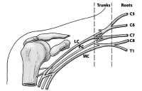
The basic anatomical relationships of the brachial plexus (BP). The BP is subdivided into roots, trunks, divisions, cords, and branches. LC stands for lateral cord, PC stands for the posterior cord, and MC stands for the medial cord
The BP supplies all of the cutaneous innervation of the upper limb, except for the area of the axilla (which is supplied by the supraclavicular nerve) and the dorsal scapula area, which is supplied by cutaneous branches of the dorsal rami.

The BP communicates with the sympathetic trunk via gray rami communicantes, which join the roots of the plexus. They are derived from the middle and inferior cervical sympathetic ganglia and the first thoracic sympathetic ganglion.

Gross Anatomy
Brachial plexus architecture
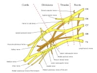
The brachial plexus (BP) is subdivided into roots, trunks, divisions, cords, and branches. Several mnemonics can be used to remember this architecture (eg, Really Tired Drink Coffee Black). Typically, the brachial plexus is composed of 5 roots, 3 trunks, 6 divisions, 3 cords, and terminal branches, as seen in the image below
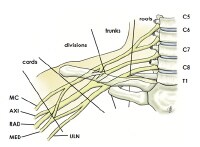
Brachial plexus with terminal branches labeled. MC is musculocutaneous (nerve), AXI is axillary, RAD is radial, MED is median, and ULN is ulnar.

Roots
The ventral rami of spinal nerves C5 to T1 are referred to as the "roots" of the plexus. The typical spinal nerve root results from the confluence of the ventral nerve rootlets originating in the anterior horn cells of the spinal cord and the dorsal nerve rootlets that join the spinal ganglion in the region of the intervertebral foramen.

The roots emerge from the transverse processes of the cervical vertebrae immediately posterior to the vertebral artery, which travels in a cephalocaudad direction through the transverse foramina. Each transverse process consists of a posterior and anterior tubercle, which meet laterally to form a costotransverse bar. The transverse foramen lies medial to the costotransverse bar and between the posterior and anterior tubercles. The spinal nerves that form the brachial plexus run in an inferior and anterior direction within the sulci formed by these structures.

Trunks
Shortly after emerging from the intervertebral foramina, the 5 roots (C5-T1) unite to form 3 trunks. The trunks of the BP pass between the anterior and middle scalene muscles.

The ventral rami of C5 and C6 unite to form the upper trunk. The suprascapular nerve and the nerve to the subclavius arise from the upper trunk. The suprascapular nerve contributes sensory fibers to the shoulder joint and provides motor innervation to the supraspinatus and infraspinatus muscles.

The ventral ramus of C7 continues as the middle trunk. The ventral rami of C8 and T1 unite to form the lower trunk .

Divisions
Each trunk splits into an anterior division and a posterior division. These separate the innervation of the ventral and dorsal aspect of the upper limb. The anterior divisions usually supply flexor muscles. The posterior divisions usually supply extensor muscles.

Cords
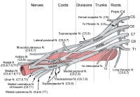
The cords are referred to as the lateral, posterior, and medial cord, according to their relationship with the axillary artery, as seen in the image below.


The cords pass over the first rib close to the dome of the lung and continue under the clavicle immediately posterior to the subclavian artery.

The anterior divisions of the upper and middle trunks unite to form the lateral cord, which is the origin of the lateral pectoral nerve (C5, C6, C7).

The anterior division of the lower trunk forms the medial cord, which gives off the medial pectoral nerve (C8, T1), the medial brachial cutaneous nerve (T1), and the medial antebrachial cutaneous nerve (C8, T1).

The posterior divisions from each of the 3 trunks unite to form the posterior cord.

The upper and lower subscapular nerves (C7, C8 and C5, C6, respectively) leave the posterior cord and descend behind the axillary artery to supply the subscapularis and teres major muscles. The thoracodorsal nerve to the latissimus dorsi (also known as the middle subscapular nerve, C6, C7, C8) also arises from the posterior cord, as seen in the image below.
Musculocutaneous nerve branch
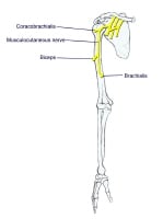
These are mixed nerves containing both sensory and motor axons (as seen in the image below). The musculocutaneous nerve is derived from the lateral cord. The musculocutaneous nerve leaves the BP sheath high in the axilla at the level of the lower border of the teres major muscle and passes into the coracobrachialis muscle. It innervates the muscles in the flexor compartment of the arm. It carries sensation from the lateral (radial) side of the forearm.
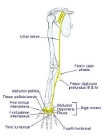
Ulnar nerve branch
The ulnar nerve is derived from the medial cord. Motor innervation is mainly to intrinsic muscles of the hand (as seen in the image below). Sensory innervation is from the medial (ulnar) 1.5 digits (little finger and one-half of the ring finger).
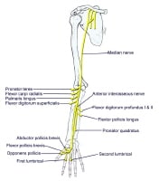
Median nerve branch
The median nerve is derived from both the lateral and medial cords. Motor innervation is to most flexor muscles in the forearm and intrinsic muscles of the thumb (thenar muscles), as seen in the image below. Sensory innervation is to the lateral (radial) 3.5 digits (thumb, index, middle, and half of the ring finger).
Axillary nerve branch
The axillary nerve is derived from the posterior cord. The axillary nerve leaves the BP at the lower border of the subscapularis muscle and continues along the inferior and posterior surface of the axillary artery as the radial nerve. The axillary nerve serves as motor innervation to the deltoid and teres minor muscles, as seen in the image below. These act at the glenohumeral joint. Sensory innervation is from the skin just below the point of the shoulder. The axillary nerve continues as the superior lateral brachial cutaneous nerve of the arm.
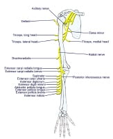
Radial nerve branch
The radial nerve is also derived from the posterior cord. The radial nerve continues along the posterior and inferior surface of the axillary artery. The radial nerve innervates the extensor muscles of the elbow, wrist, and fingers, as seen in the image above. Sensory innervation is from the skin on the dorsum of the hand on the radial side.

Terminal Branches
Five "terminal" branches and numerous other "pre-terminal" or "collateral" branches leave the plexus at various points along its length.

Dorsal scapular nerve
The dorsal scapular nerve is derived from the C5 root just after its exit from the intervertebral foramen. It serves as the motor nerve to the rhomboids major and minor muscles

Long thoracic nerve
The long thoracic nerve is derived from C5, C6, and C7 roots immediately after their emergence from the intervertebral foramina. The long thoracic nerve crosses the first rib and then descends through the axilla behind the major branches of the plexus. It innervates the serratus anterior muscle.

Phrenic nerve
The phrenic nerve arises from C3, C4, and C5 root levels, chiefly from the C4 nerve root. It crosses the anterior scalene from lateral to medial and extends into the thorax between the subclavian vein and artery.

Subclavius muscle nerve
The nerve to the subclavius muscle is a small filament, which arises from the upper trunk. It descends to the subclavius muscle in front of the subclavian artery and the lower trunk of the plexus.

Suprascapular nerve
The suprascapular nerve arises from the upper trunk formed by the union of the fifth and sixth cervical nerves. It innervates the supraspinatus muscles and infraspinatus muscles. It runs laterally beneath the trapezius and the omohyoideus and enters the supraspinatus fossa through the suprascapular notch, below the superior transverse scapular ligament; it then passes beneath the supraspinatus and curves around the lateral border of the spine of the scapula to the infraspinatus fossa.

Lateral pectoral nerve
The lateral pectoral nerve arises from the lateral cord of the brachial plexus, from the fifth, sixth, and seventh cervical nerves. It passes across the axillary artery and vein, pierces the coracoclavicular fascia, and is distributed to the deep surface of the pectoralis major. It sends a filament to join the medial anterior thoracic and forms with it a loop in front of the first part of the axillary artery. This nerve innervates the clavicular head of the pectoralis major muscle.

Medial pectoral nerve
The medial pectoral nerve arises from the medial cord from the eighth cervical and first thoracic nerve. It passes behind the first part of the axillary artery, curves forward between the axillary artery and vein, and unites in front of the artery with a filament from the lateral nerve. It then enters the deep surface of the pectoralis minor, where it divides into a number of branches, which supply the muscle. Several branches of the medial pectoral nerve pierce the muscle and end in the pectoralis major, which supply the muscle.

The medial and lateral pectoral nerve often join together to act as a single nerve innervating both the pectoralis major and minor muscles.

Medial brachial cutaneous nerve
The medial brachial cutaneous nerve is the smallest branch of the brachial plexus and, arising from the medial cord, receives its fibers from the eighth cervical and first thoracic nerves. It passes through the axilla, at first lying behind and then medial to the axillary vein, and communicates with the intercostobrachial nerve. It descends along the medial side of the brachial artery to the middle of the arm, where it pierces the deep fascia, and is distributed to the skin of the back of the lower third of the arm, extending as far as the elbow, where some filaments are lost in the skin in front of the medial epicondyle, and others over the olecranon. It communicates with the ulnar branch of the medial antebrachial cutaneous nerve. It carries sensation from the lower medial portion of the arm.

Medial antebrachial cutaneous nerve
The medial antebrachial cutaneous arises from the medial cord of the brachial plexus. It derives its fibers from the eighth cervical and first thoracic nerves, and at its commencement is medial to the axillary artery. It gives off, near the axilla, a filament, which pierces the fascia and supplies the integument covering the biceps brachii, nearly as far as the elbow. The nerve then runs down the ulnar side of the arm medial to the brachial artery, pierces the deep fascia with the basilic vein, about the middle of the arm, and divides into a volar and an ulnar branch.

Microscopic Anatomy
Blood supply of the brachial plexus
The blood supply of the brachial plexus is based largely on the subclavian (which becomes the axillary) artery and its branches, and variations exist. Generally, the vessels involved are the vertebral, the ascending and deep cervical, and the superior intercostal arteries. The cord and rootlets of the cervical nerves are supplied by the anterior and posterior spinal branches of the vertebral artery. The trunks of the plexus are supplied by muscular branches of the ascending and deep cervical arteries and superior intercostals, and occasionally from the subclavian itself.

Natural Variants
Many variant forms of the brachial plexus exist, with none representing most patients. Kerr catalogued 29 forms of the brachial plexus among some 175 cadaver specimens dissected between 1895 and 1910.[2] In the early part of the last century, one author described a total of 38 variations of the plexus. Up to 53.5% of plexuses in cadaver studies possess significant anatomic variation from the "classic" description of the brachial plexus. A study published in September 2003 found variants in 107 of 200 fetuses examined. The authors pointed out that morphological variations were more common in female fetuses and right sides.[3]

Prefixed brachial plexus (cephalic or high)
This occurs when the C4 ventral ramus contributes to the brachial plexus. Contributions to the plexus usually come from the C4-C8.

Postfixed brachial plexus (caudal or low)
This occurs when the T2 ventral ramus contributes to the brachial plexus. Contributions to the plexus usually come from C6-T2.

Other Considerations
Lesions of the Brachial Plexus
Knowledge of the muscles innervated by branches of the brachial plexus and the actions of these muscles and areas of anesthesia and/or paraesthesia allows the physician to determine the localization of a given lesion.

Brachial plexus injuries have numerous causes, such as labor and delivery. A plexus injury at birth is likely caused by a stretch or tear of the child's brachial plexus during the delivery process. The abducted arm of the infant can get pinned against the child's head. This injury results in incomplete sensory and/or motor function of the injured nerve. Traumatic brachial plexus injuries may occur due to motor vehicle accidents, bike accidents, ATV accidents, or sports.

Nerve injuries vary in severity from a mild stretch to the nerve root tearing away from the spinal cord.

Avulsion
The nerve is torn away from its attachment at the spinal cord; this is the most severe type of injury.

Rupture
The nerve is torn, but not at the spinal cord attachment.

Neuroma
Scar tissue has grown around the injury site, putting pressure on the injured nerve and preventing the nerve from sending signals to the muscles.

Neurapraxia
The nerve has been stretched and damaged but not torn.

Diagnoses Related to Brachial Plexus Injuries
Diagnoses related to brachial plexus injuries include Erb palsy, Klumpke palsy, thoracic outlet syndrome, Burner syndrome, and Parsonage-Turner syndrome.

Erb palsy
This condition involves the upper root nerves of C5, C6.
The patient often presents with the arm extended and wrist fully flexed (waiter's tip).
Motor deficits are loss of abduction, flexion, and rotation at the shoulder (axillary, suprascapular, upper, and lower subscapular nerve); weak shoulder extension; and weak elbow flexion and supination of the radioulnar joint (musculocutaneous and radial nerve).
The patient is susceptible to shoulder dislocation due to loss of rotator cuff muscles .
Sensory deficits are to the posterior and lateral aspect of arm (axillary nerve ), radial side of the forearm (musculocutaneous nerve), and thumb and first finger (superficial branch of radial nerve; digital branches of the median nerve ).

Klumpke palsy
This condition involves the lower root nerves of C8 and T1.
This is caused by a rare injury of the lower brachial plexus, usually following breech delivery.
Motor deficits are opposite the thumb (thenar branch of the median nerve), loss of adduction of the thumb (ulnar nerve), loss of abduction and adduction of metatarsophalangeal joints; flexion at the metatarsophalangeal and extension of the interphalangeal joints (deep branch of ulnar and median), and weak flexion of proximal interphalangeal joints and distal interphalangeal joints (ulnar and median nerve).
Sensory deficit is to the ulnar side of the forearm, hand, and ulnar one and a half digits (ulnar and medial antebrachial cutaneous nerve).

Thoracic outlet syndrome
This condition is caused by compression of the neurovascular structures (subclavian vessels, brachial plexus, cervical ganglia, and vertebral artery) in the cervicoaxillary region. It may be congenital or acquired.
Bony causes include the following: long transverse process of the seventh cervical vertebra, cervical rib, anomalous first rib, clavicle fracture, and first rib fracture.
Soft tissue causes include the following: congenital bands, poor posture, mass lesions, cervical strain, hypertrophy, and/or injury of the anterior and middle scalene muscles or the pectoralis minor muscle. 

Burner syndrome
This condition is thought to be related to brachial plexus stretch and/or nerve root compression. It generally involves the upper trunk of the brachial plexus, particularly C5-C6.
This condition is usually a result of traction injury with axial distraction or downward force on the shoulder combined with lateral bend of the neck away from the shoulder.
Brachial plexus stretch injuries are found in young athletes, with neck pain being less common.
Nerve root compression is found in older athletes (college), generally associated with cervical disc disease or cervical stenosis. Neck pain is more common. 

Parsonage-Turner syndrome (brachial neuritis)
This condition is generally associated with a viral prodrome, immunizations, and significant pain.
Acute excruciating unilateral shoulder pain is present, followed by flaccid paralysis of shoulder and parascapular muscles several days later.
This condition varies greatly in manifestation and nerve involvement. Due to the extreme pain involved, patients usually present acutely. Often, the affected arm is supported by the uninvolved arm and is held in adduction and internal rotation.
The patient may have atrophy of the affected muscles.
Pain may occur with palpation and active and passive range of motion.
Reflexes may be reduced/absent, depending on the nerves involved.
Sensory loss is usually not prominent, although it may be detectable.
This condition may cause scapular winging.